Paper and Paper Microfluidics for Analytical Devices and Sensors
ABSTRACT
1. Introduction
Paper is a well-known material to record information for handwriting and printing. Nowadays, paper is used in various applications, after appropriate modifications and engineering. For example, it can be used for packing and storing applications,1,2) for packaging food and drink,3,4) as an absorber of physical shocks,5,6) as a heat and electromagnetic-wave insulator,7,8) as a shield for oscillating sounds,9) for filtration,10) and in many other applications. In recent years, paper has also become a popular material for the research and development of analytical devices and sensors.
Sensors are commonly developed using electrical,11) optical, or spectral12-14) imaging15-17) technology to identify and/or quantify properties of interest. However, when a biological and/or chemical reaction during a measurement is required, a sensor spatial area and volume are needed for the reaction to occur. Typically, a well-plate is commonly used in many assays like enzyme-linked immunosorbent assay. The assay requires multiple stages of washing and reaction processes in each well, and the whole plate is taken to a specifically developed reader, in order to obtain the result from each well.18) The use of well-plates requires a tedious and handful process at each stage during complex procedures. A microfluidic chip system is introduced during the sensor’s research and development stages in order to overcome the difficulties associated with the use of well-plates. The microfluidic chip is fabricated with transparent polymers (i.e., polydimethylsiloxane or polymethylmethacrylate) that have patterned channels to allow liquid samples to flow through. Micro-pumps deliver multiple samples into the microfluidic chip during the reaction.19,20) The system requires a small number of samples and can become automated by programming the operation of the micro-pumps. However, the cost of such a system is high because of a) the etching process on a silicon wafer to generate the patterned mold for the polymer chip and b) the cost of purchasing the micro-pump. On the other hand, paper has become an alternative material with increasing interest in the microfluidic apparatus for the development of analytical devices and sensors.
Paper-based analytical devices or sensors have several advantages because they are cost-effective, easy to carry, and disposable; they do not require filters for purifying the samples or pumping apparatuses to deliver the liquid sample. These advantages are based on the cellulose fibers, which form the porous structure of the paper. The porous structure which filters out macro-size molecules in the sample, are mostly irrelevant to the material of interest. The pores (with a size of <100 μm) create a capillary structure for the liquid sample to flow through. Also, the cellulose fiber has a high affinity with hydrophilic substances due to the abundance of the hydroxyl group in the cellulose molecules, leading to strong wetting and capillary effect with hydrophilic liquids. Moreover, biomolecules can be covalently bound to the surface of the cellulose. In addition, due to the high surface-to-volume ratio, these biomolecules can be efficiently covered on the cellulose surface and enhance the signal-to-noise ratio of the sensor.
Paper has become a very popular material in the field due to the introduction of a hydrophobic material patterning technique on the paper. There are two major techniques of patterning hydrophobic barrier on paper: a) photolithography21) and b) wax printing.22) Easier paper patterning techniques have resulted in its application not only in the analytical device and sensors but also in numerous other fields, such as electronics and biology. The pattern guides the liquid flow to generate separation and mixing in order to control the liquid flow on the paper. This patterned-paper platform is called paper microfluidics and has been applied to the development of sensors for various applications, such as clinical, pharmaceutical, environmental, agricultural, and food applications.
In this paper, we analyze research trends by investigating a number of scientific articles related to paper microfluidics and the application of paper microfluidics to the sensors’ development. General information related to the commonly used paper patterning methods for the fabrication of paper microfluidics is presented. Moreover, representative research cases of using the paper microfluidics in the development of sensors are described.
2. Research Trends in Paper Microfluidics
The hydrophobic patterning technique introduced in 2007 enabled paper to contain a liquid sample or a mix of different liquid samples in a controlled manner.21) Since then, the number of articles related to paper research has drastically increased in the science and engineering field. Two-dimensional,23) three-dimensional flow24) paper microfluidics, and even paper origami25,26) were investigated for various purposes.
Statistical data were produced by analyzing the number of studies related to paper and paper microfluidics for various research purposes for the last 10 years since 2008. The number of the published articles has continuously increased, and the largest number of papers was published last year, 2018. Most of these publications are related to paper or paper microfluidics with respect to the development of analytical devices and sensors (Fig. 1). Other research studies on paper presented applications, such as paper-based battery,27,28) electrical circuit on paper boards and flexible electronics,29,30) paper-based antennas,31,32) RFIDs,33) capacitive touch pads,34) and cell culture.35,36) Almost 80% of the research articles are related to the use of paper for the development of sensors. This is due to the attractive properties of paper as a material for sensor development. A paper-based sensor is relatively cheap, easy to carry, easy-to-use, and disposable; therefore, it fulfills the demand of sensor being the point-of-use. The number of research papers related to paper microfluidics is constantly increasing and paper microfluidics has gained popularity.
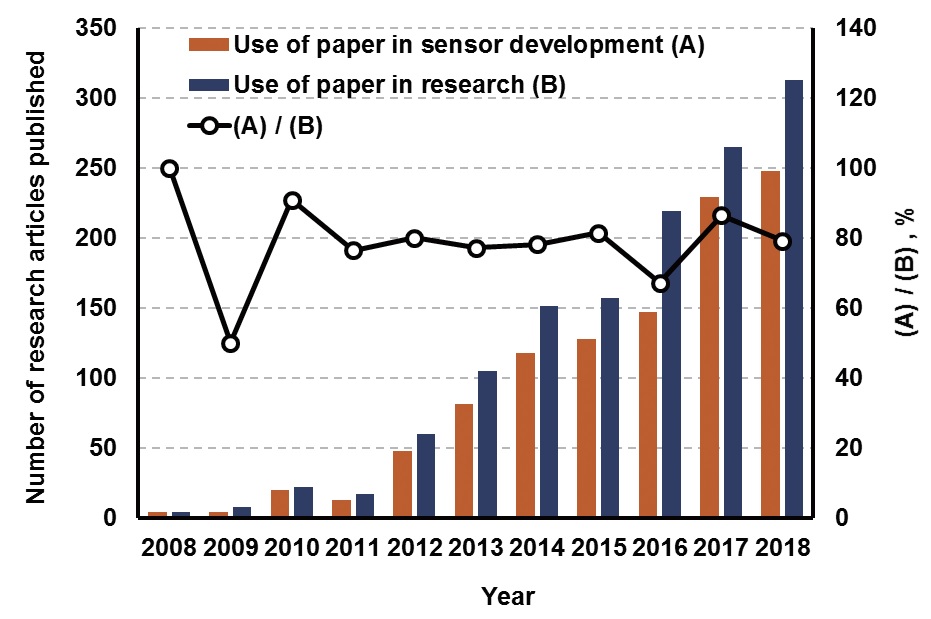
Fig. 1.
Number of articles published that are related to paper research in general and to sensor development using paper in particular, during the last 10 years. Data were mined from the Web of Science using the following keywords: “Paper-based,” “Paper microfluidics,” and “Microfluidic paper-based analytical device.”
3. Hydrophobic Paper Patterning Methods
3.1 Wax printing method
In the wax printing method, the wax printer is used to print wax on paper with a custom designed pattern. The wax is a hydrophobic substance that can create a barrier and a guide for a hydrophilic sample to flow through the hydrophilic region of the paper. Wax printing method requires two simple steps to create a pattern on the paper. In the first step, the pattern is printed on paper using the wax printer, and in the second step wax is melted to fill the entire paper layer (Fig. 2).
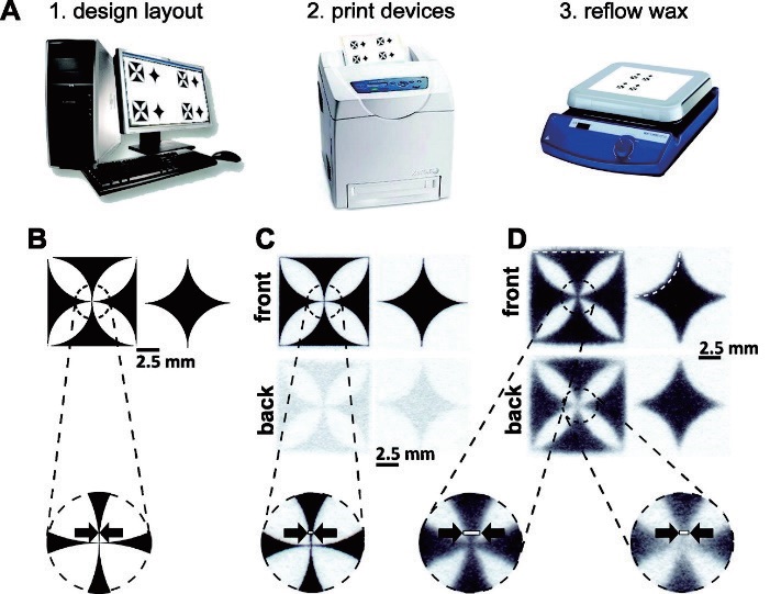
Fig. 2.
Overview of the patterning process using a wax printer (A). A pattern is created using design programs (B). Shape of the printed wax on paper before the melting process and details of the front and back of the paper from which the pattern was printed (C). Front and back of the paper with blurred wax (D)22) after the heating process.
The wax printing method has a limitation that relates to limited pattern accuracy. This is because printed wax above the paper diffuses in all directions through the fibers in the paper during the heating process (Fig. 2B). The blurred pattern created after heating may cause a change in the flow rate in the channel and in the amount of sample to be introduced for analysis. However, this patterning method has the great advantage of being a faster process with bulk production method compared with the photolithography method.
3.2 Photolithography method
In the photolithography method, a light-sensitive material (photoresist) is applied on the paper and covers it with a patterned photomask during the UV exposure. The residue of the photoresist is removed (Fig. 3). The remained photoresist pattern on the paper functions as a barrier and guides the liquid sample to flow within the cellulose fibers of the paper. The photolithography method is a reproducible and sophisticated method suitable for developing a pattern on the paper. However, it has disadvantages, such as the use of materials and instruments and requires a longer time to create a pattern.

Fig. 3.
(A) A brief description of the patterning process using a photoresist. (B) Fabrication of the photomask of a designed pattern. (C) UV light is radiated on the paper that is impregnated with the photomask and photoresist. (D) After the irradiation of UV light, the photomask is removed from the paper. (E) Application of heating to bake the paper. (F) After the application of the washing process using the organic solvent, the completed paper-based microfluidic chip21) is created. (G) SEM image of the border between the paper channel (left) and the photoresist layer (right), modified from Park et al.37)
4. Analytical Device and Sensor Applications Using Paper Microfluidics
The applications of paper-based analytical devices and sensors can be classified into four categories: a) point-of-care (POC) testing in biomedical engineering, b) food safety, c) agriculture, and d) environmental monitoring. The detection of the material of interest is accomplished using mostly optical and electrochemical detection methods. Measurements of color change38-40) or the fluorescence signal41,42) from reactions are the most common of the optical methods in paper microfluidics. Recently, these optical detections from the paper microfluidics were measured and analyzed by a smartphone camera and an app.37,43)
Electrochemical detection requires electrodes on paper microfluidics for potentiometric44,45) and amperometric46,47) measurements during oxidation/reduction between the sample and the electrodes. Carbon nanotubes,48) carbon tape,49) graphene,46) and pencil drawings50) were used to create the electrodes in the paper microfluidics.
4.1 Paper microfluidics for POC devices
The POC testing is an advanced method for the immediate medical treatment of patients on the site and typically includes blood glucose level, urine, and tumor marker tests. A three-dimensional paper-based microfluidic chip was developed for detecting tumor markers that can identify the presence of cancer cells at an early stage.51) Tumor marker testing is a type of cancer test that uses blood as a sample. The tumor markers include prostate-specific antigen, r-fetoprotein (AFP), carcinoma antigen 125 (CA125), carcinoma antigen 199 (CA199), carcinoma antigen (CA15-3), and carcinoembryonic antigen (CEA), four tumor markers (AFP, CA125, CA199, and CEA) were analyzed using paper microfluidics (Fig. 4). A sample that is dropped on the center circle of the paper microfluidics flows through each of the six channels. Tumor markers in the sample bind with antibody that is immobilized on each detection site. A second antibody that is pre-bound with Ru-labeled binds to the tumor marker creates a sandwich binding that generates the electrochemiluminescence signal to quantify the amount of tumor present in the sample.
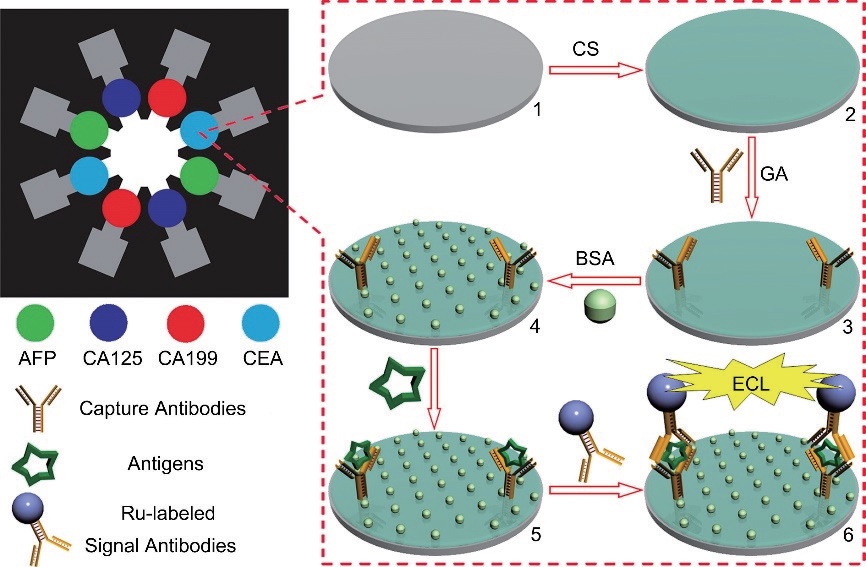
Fig. 4.
Schematic diagram of the analysis procedures and three-dimensional paper-based microfluidic chips for the detection of tumor markers. An antibody that causes a specific response to a particular tumor marker is immobilized on the surface of the carbon working electrode.51)
Tumor markers were detected using an electrochemiluminescence method that combines the advantages of chemiluminescence and electrochemistry. Two working electrodes are placed per each tumor marker. Four standard curves of each tumor marker were created. All correlation coefficients have values of 0.99 or higher (R=0.9978 (AFP), R=0.9971 (CA125), R=0.9968 (CA199), and R=0.9963 (CEA)). The limit of detection for each tumor marker was as follows: 0.15 ng/mL (AFP), 0.6 U/mL (CA125), 0.17 U/mL (CA199), and 0.5 ng/mL (CEA). Alternatively, the sensor of the tumor marker CEA using an indium tin oxide electrode with chitosan was produced, without using paper as a material.52) The CEA was detected using the electrochemiluminescence method. The standard curve created indicated a detection range of 0.005-200 ng/mL and a correlation coefficient value of 0.998. Compared with three-dimensional paper-based microfluidic chips, they exhibit a wider limit of detection and detection range. Considering that the cutoff value of the CEA tumor marker is 5 ng/mL in the actual clinical diagnosis, the performance of the tumor marker detection sensor when paper is used is also sufficiently meaningful.53)
4.2 Paper microfluidics for food safety analysis
In the field of food safety, the main point of interest is to detect microorganisms and toxins that cause food poisoning. A paper-based sensor using the colorimetry method was developed to detect a food-borne pathogen.54)Escherichia coli O157:H7, Listeria monocytogenes, and Salmonella Typhimurium, which are typical pathogens causing food poisoning, were detected in this research work. In order to determine the concentration of pathogens, the colors produced by specific reactions between enzymes and substrates by each microorganism were analyzed. Esterase with 5-bromo-6-chloro-3indolyl caprylate (magenta caprylate) reaction, β-galactosidase with chlorophenol red β-galactopyranoside (CPRG) reaction, and phosphatidylinositol-specific phospholipase C (PI-PLC) with 5-bromo-4-chloro-3indolyl-myo-inositol phosphate (X-InP) reaction were used for the detection of Salmonella Typhimurium, E. coli O157:H7, and L. monocytogenes, respectively.
Ready-to-eat meat products were inoculated with bacteria and cultured sufficiently. The samples were used as extracts with bacterial concentration values of 101, 102, and 103 CFU/cm2. The paper-based sensor was able to detect bacteria at a concentration of 101 CFU/cm2 after they were enriched for about 8 to 12 h in media (Fig. 5). Although the detection range is limited, this method has the advantage of time saving when compared with the conventional plate culture method. Additionally, bacteria can be detected from real food, and there is no need for any additional equipment to be used in the analysis. Moreover, there is no need of a biosensor that reacts with specific pathogens or a separate filtration stage to remove foreign substances from actual extracts. The porous structure of the paper helps to filter out substances larger than the pore size except for the biomolecules to be analyzed.
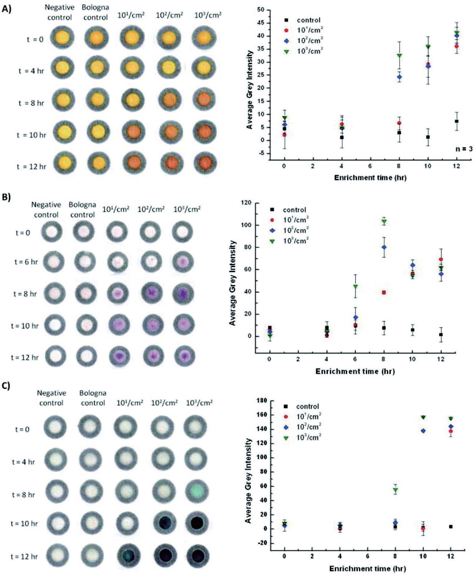
Fig. 5.
After inoculating pathogens into ready-to-eat meat products, the extracts were used as samples and analyzed by applying them to a paper-based microfluidic chip (A) E. coli O157:H7, (B) Salmonella Typhimurium, and (C) L. monocytogenes.54)
Another method for detecting food-borne pathogens is the combination of paper microfluidics with a smartphone camera. The surface of the sub-micron size poly-styrene particle was immobilized and covered by the antibody for Salmonella Typ. The particle was preloaded in the middle of the paper channel ready to bind and form aggregates with the introduction of bacteria. The particle exhibits different light scatter characteristics depending on the degree of aggregation that corresponds to the amount of target bacteria present in the sample. The difference in light scattering before and after the particle aggregation was measured by a smartphone camera, and the result of Salmonella in the sample was printed out (Fig. 6).37)
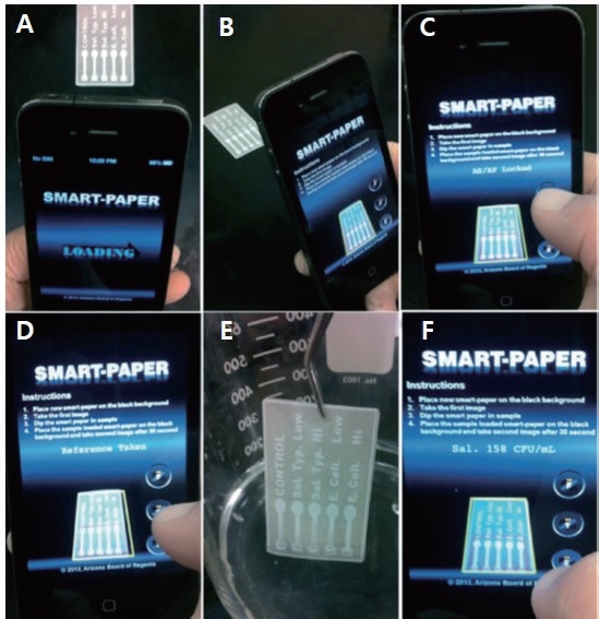
Fig. 6.
Images of the smartphone workflow application to detect Salmonella on multi-channel paper microfluidics. Smartphone loads the developed app (A); the app guides the user to adjust the angle and the distance between the camera and the paper microfluidics (B), locking auto exposure and auto focus of the camera (C), taking a reference image with dried Ab-PS loaded on the paper microfluidics (D), sample loading on the paper microfluidics (E), and displaying the assay result after taking the signal image of the paper microfluidics (F).37)
4.3 Paper microfluidics for agricultural and environmental applications
Measuring the amount of proline in a plant's leaf helps to analyze and diagnose the degree of drought stress applied to the plant. The detection and estimation of the amount of proline can be done in a fast and simple way, using a three-dimensional origami fabricated paper microfluidic (Fig. 7). Proline–ninhydrin reaction is executed in the paper microfluidics, and the reagent required for the reaction is loaded on the sensing part of the sensor. After the sample from a plant leaf is dropped on the sensing area, the reaction occurs and a red color is developed. The color intensity is measured and analyzed to diagnose the early stage of drought stress on the plant.55)
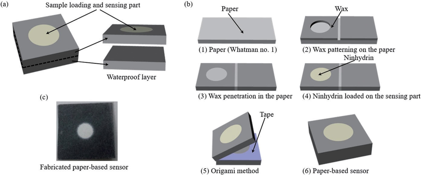
Fig. 7.
Schematic diagram of (a), manufacturing process (b), fabricated paper-based sensor for drought stress diagnosis in plants (c).55)
Environmental monitoring is mainly the detection of toxins or heavy metals that cause soil and water pollution. Gold nanoparticles that are immobilized with single-stranded DNA (ssDNA) on the surface are spread on paper microfluidics to detect mercury(II) ions.56) If mercury(II) ions exist in the sample, the ssDNA forms Thymine-Hg2+- Thymine bonds and then loses its binding affinity with the nanoparticle surfaces. At this moment, aggregation occurs between nanoparticles without ssDNA on the surface, and the color changes depending on the degree of aggregation. The concentration of mercury(II) ions is determined by analyzing the degree of the color change (Fig. 8). Samples are collected from actual ponds and rivers and used to create the standard curve. The detection range of the developed standard curve is 25-100 nM, and the limit of detection is about 50 nM.
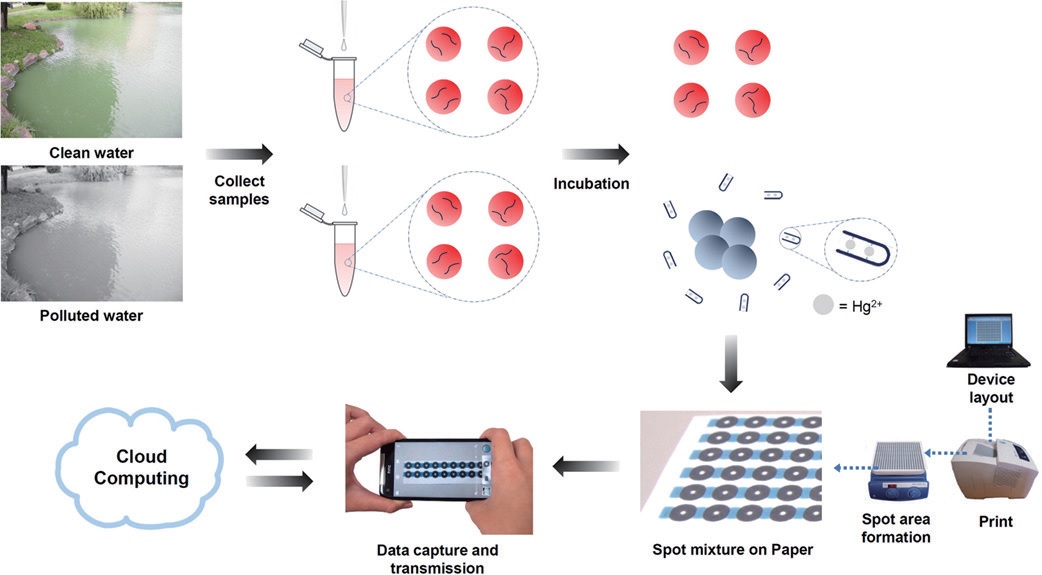
Fig. 8.
Schematic diagram representing the process from actual sampling to application into paper-based microfluidic chip.56)
5. Conclusions
In this article, the use of paper as a sensor material for the development of analytical devices and sensors was analyzed and presented. Paper can be applied to develop POC diagnosis and analytical devices and sensors for food-borne pathogens and for agricultural and environmental purposes. It has been proved that paper is a promising sensor material that has many advantages. First, no additional apparatus such as pumps are required to transport or deliver the sample due to the capillary effect of paper. Second, paper can provide scaffolds capable of immobilizing a substrate that has a specific binding to a particular biomolecule and channels to transport a sample. Third, simple and reproducible patterning technology makes it easy to fabricate patterns on paper. Finally, since the paper is composed of cellulose fiber, it is easily disposable for the single-use sensor. Because of these merits of using paper in the sensor development, it is expected that research on paper applications will significantly grow and gain popularity.
Acknowledgements
This research was supported by the Kyungpook National University Research Fund, 2016.
Literature Cited
C. K. Min, J. Y. Jo and J. S. Shin, Journal of Korea TAPPI,
The effect of the application of the acid liquid from rice hull carbonization to molded fiber packages on the shelf life of egg,
50(6); 108-114 (2018)
Min, C. K., Jo, J. Y., and Shin, J. S., The effect of the application of the acid liquid from rice hull carbonization to molded fiber packages on the shelf life of egg, Journal of Korea TAPPI 50(6):108-114 (2018).
10.7584/jktappi.2018.12.50.6.108D. W. Jang and J. M. Park, Journal of Korea TAPPI,
Changes of handsheet’s physical properties depending on mixing ratio of bagasse pulp for replacement of hardwood bleached kraft pulp,
49(1); 3-8 (2017)
Jang, D. W. and Park, J. M., Changes of handsheet’s physical properties depending on mixing ratio of bagasse pulp for replacement of hardwood bleached kraft pulp, Journal of Korea TAPPI 49(1):3-8 (2017).
10.7584/jktappi.2017.02.49.1.3G. Kyung, H. Yang, W. Lee, J. Park and S. Ko, Journal of Korea TAPPI,
Evaluation of the antibacterial and physical properties of paper coated with chitosan-Ag nanocomposite prepared by green synthesis,
46(4); 28-36 (2014)
Kyung, G., Yang, H., Lee, W., Park, J., and Ko, S., Evaluation of the antibacterial and physical properties of paper coated with chitosan-Ag nanocomposite prepared by green synthesis, Journal of Korea TAPPI 46(4):28-36 (2014).
10.7584/ktappi.2014.46.4.028S. R. Back, L. Melani and H. -J. Kim, Journal of Korea TAPPI,
Physical properties and antimicrobial behaviour of food packaging paper by green tea extracts,
49(6); 5-12 (2017)
Back, S. R., Melani, L., and Kim, H. -J., Physical properties and antimicrobial behaviour of food packaging paper by green tea extracts, Journal of Korea TAPPI 49(6):5-12 (2017).
10.7584/jktappi.2017.12.49.6.5Y. -M. Lee, C. -H. Kim, J. -O. Kim, G. -Y. Kim, T. -G. Shin, D. B. Song and C. -Y. Park, Journal of Korea TAPPI,
Development of environment-friendly cushioning materials by pulping of waste residual woods,
38(2); 61-71 (2006)
Lee, Y. -M., Kim, C. -H., Kim, J. -O., Kim, G. -Y., Shin, T. -G., Song, D. B., and Park, C. -Y., Development of environment-friendly cushioning materials by pulping of waste residual woods, Journal of Korea TAPPI 38(2):61-71 (2006).
G. -Y. Kim, C. -H. Kim, Y. -M. Lee, D. -B. Song, T. -G. Shin, J. -O. Kim and C. -Y. Park, Journal of Korea TAPPI,
Utilization of wastepaper fibers for development of environment-friendly shock-absorbing materials,
38(2); 52-60 (2006)
Kim, G. -Y., Kim, C. -H., Lee, Y. -M., Song, D. -B., Shin, T. -G., Kim, J. -O., and Park, C. -Y., Utilization of wastepaper fibers for development of environment-friendly shock-absorbing materials, Journal of Korea TAPPI 38(2):52-60 (2006).
Y. J. Sung, M. H. Heo, H. S. Chung and J. Y. Lee, Journal of Korea TAPPI,
Effects of the structure and the inorganic filler type on the heat insulation of paper,
46(4); 93-100 (2014)
Sung, Y. J., Heo, M. H., Chung, H. S., and Lee, J. Y., Effects of the structure and the inorganic filler type on the heat insulation of paper, Journal of Korea TAPPI 46(4):93-100 (2014).
10.7584/ktappi.2014.46.4.093J. Y. Lee, C. H. Kim, H. G. Nam, H. H. Park, S. Kwon and Y. M. Lee, Journal of Korea TAPPI,
Development of functional pulp tray for prevention of static electricity,
47(5); 52-60 (2015)
Lee, J. Y., Kim, C. H., Nam, H. G., Park, H. H., Kwon, S., and Lee, Y. M., Development of functional pulp tray for prevention of static electricity, Journal of Korea TAPPI 47(5):52-60 (2015).
10.7584/ktappi.2015.47.5.052C. W. Kang and Y. B. Seo, Journal of Korea TAPPI,
Sound absorption and sound transmission loss of perforated corrugated board,
50(4); 32-39 (2018)
Kang, C. W. and Seo Y. B., Sound absorption and sound transmission loss of perforated corrugated board, Journal of Korea TAPPI 50(4):32-39 (2018).
10.7584/jktappi.2018.08.50.4.32S. Heydarifard, K. Taneja, G. Bhanjana, N. Dilbaghi, M. M. Nazhad, K. H. Kim and S. Kumar, Carbohydrate Polymers,
Modification of cellulose foam paper for use as a high-quality biocide disinfectant filter for drinking water,
181; 1086-1092 (2018)
Heydarifard, S., Taneja, K., Bhanjana, G., Dilbaghi, N., Nazhad, M. M., Kim, K. H., and Kumar, S., Modification of cellulose foam paper for use as a high-quality biocide disinfectant filter for drinking water, Carbohydrate Polymers 181:1086-1092 (2018).
10.1016/j.carbpol.2017.11.038D. W. Kim, D. H. Jung, W. J. Cho, K. C. Sim and H. J. Kim, Journal of Biosystems Engineering,
On-site water nitrate monitoring system based on automatic sampling and direct measurement with ion-selective electrodes,
42(4); 350-357 (2017)
Kim, D. W., Jung, D. H., Cho, W. J., Sim, K. C., and Kim, H. J., On-site water nitrate monitoring system based on automatic sampling and direct measurement with ion-selective electrodes, Journal of Biosystems Engineering 42(4):350-357 (2017).
S. J. Hong, A. Y. Lee, Y. H. Han, J. Park, J. D. So and G. Kim, Journal of Biosystems Engineering,
Rancidity prediction of soybean oil by using near-infrared spectroscopy techniques,
43(3); 219-228 (2018)
Hong, S. J., Lee, A. Y., Han, Y. H., Park J., So, J. D., and Kim, G., Rancidity prediction of soybean oil by using near-infrared spectroscopy techniques, Journal of Biosystems Engineering 43(3):219-228 (2018).
J. G. Lim, G. Y. Kim, C. Y. Mo, K. M. Oh, G. S. Kim, H. C. Yoo, H. H. Ham, Y. T. Kim, S. M. Kim and M. S. Kim, Journal of Biosystems Engineering,
Rapid and nondestructive discrimination of Fusarium asiaticum and Fusarium graminearum in hulled barley (Hordeum vulgare L.) using near-infrared spectroscopy,
42(4); 301-313 (2017)
Lim, J. G., Kim, G. Y., Mo, C. Y., Oh, K. M., Kim, G. S., Yoo, H. C., Ham, H. H., Kim, Y. T., Kim, S. M., and Kim, M. S., Rapid and nondestructive discrimination of Fusarium asiaticum and Fusarium graminearum in hulled barley (Hordeum vulgare L.) using near-infrared spectroscopy, Journal of Biosystems Engineering 42(4):301-313 (2017).
E. Park, S. J. Hong, A. Y. Lee, J. Park, B. K. Cho and G. Kim, Journal of Biosystems Engineering,
Phenotyping of low-temperature stressed pepper seedlings using infrared thermography,
42(3); 163-169 (2017)
Park, E., Hong, S. J., Lee, A. Y., Park, J., Cho, B. K., and Kim, G., Phenotyping of low-temperature stressed pepper seedlings using infrared thermography, Journal of Biosystems Engineering 42(3):163-169 (2017).
M. R. Ahmed, J. Yasmin, W. H. Lee, C. Mo and B. K. Cho, Journal of Biosystems Engineering,
Imaging technologies for nondestructive measurement of internal properties of agricultural products: A review,
42(3); 199-216 (2017)
Ahmed, M. R., Yasmin, J., Lee, W. H., Mo, C., and Cho, B. K., Imaging technologies for nondestructive measurement of internal properties of agricultural products: A review, Journal of Biosystems Engineering 42(3):199-216 (2017).
J. Qin, M. S. Kim, K. Chao and B. K. Cho, Journal of Biosystems Engineering,
Raman chemical imaging technology for food and agricultural applications,
42(3); 170-189 (2017)
Qin, J., Kim, M. S., Chao, K., and Cho, B. K., Raman chemical imaging technology for food and agricultural applications, Journal of Biosystems Engineering 42(3):170-189 (2017).
C. Mo, J. Lim, S. W. Kwon, D. K. Lim, M. S. Kim, G. Kim, J. Kang, K. D. Kwon and B. K. Cho, Journal of Biosystems Engineering,
Hyperspectral imaging and partial least square discriminant analysis for geographical origin discrimination of white rice,
42(4); 293-300 (2017)
Mo, C., Lim, J., Kwon, S. W., Lim, D. K., Kim, M. S., Kim, G., Kang, J., Kwon, K. D., and Cho, B. K., Hyperspectral imaging and partial least square discriminant analysis for geographical origin discrimination of white rice, Journal of Biosystems Engineering 42(4):293-300 (2017).
Y. Arita, S. Kihara, N. Ouchi, M. Takahashi, K. Maeda, J. Miyagawa, K. Hotta, I. Shimomura, T. Nakamura, K. Miyaoka, H. Kuriyama, M. Nishida, S. Yamashita, K. Okubo, K. Matsubara, M. Muraguchi, Y. Ohmoto, T. Funahashi and Y. Matsuzawa, Biomedical and Biophysical Research Communications,
Paradoxical decrease of an adipose-specific protein, adiponectin, in obesity,
257(1); 79-83 (1999)
Arita, Y., Kihara, S., Ouchi N., Takahashi, M., Maeda, K., Miyagawa, J., Hotta, K., Shimomura, I., Nakamura, T., Miyaoka, K., Kuriyama, H., Nishida, M., Yamashita, S., Okubo, K., Matsubara, K., Muraguchi, M., Ohmoto, Y., Funahashi, T., and Matsuzawa, Y., Paradoxical decrease of an adipose-specific protein, adiponectin, in obesity, Biomedical and Biophysical Research Communications 257(1):79-83 (1999).
K. S. Kim and J. -K. Park, Lab on a Chip,
Magnetic force-based multiplexed immunoassay using superparamagnetic nanoparticles in microfluidic channel,
5(6); 657-664 (2005)
Kim, K. S. and Park, J. -K., Magnetic force-based multiplexed immunoassay using superparamagnetic nanoparticles in microfluidic channel, Lab on a Chip 5(6):657-664 (2005).
S. Lee, J. Choi, L. Chen, B. Park, J. B. Kyong, G. H. Seong, J. Choo, Y. Lee, K. -H. Shin, E. K. Lee, S. -W. Joo and K. -H. Lee, Analytical Chimica Acta,
Fast and sensitive trace analysis of malachite green using a surface-enhanced raman microfluidic sensor,
590(2); 139-144 (2007)
Lee. S., Choi, J., Chen, L., Park, B., Kyong, J. B., Seong, G. H., Choo, J., Lee, Y., Shin, K. -H., Lee, E. K., Joo, S. -W., and Lee, K. -H., Fast and sensitive trace analysis of malachite green using a surface-enhanced raman microfluidic sensor, Analytical Chimica Acta 590(2):139-144 (2007).
10.1016/j.aca.2007.03.049A. W. Martinez, S. T. Phillips, B. J. Wiley, M. Gupta and G. M. Whitesides, Lap on a Chip,
FLASH: A rapid method for prototyping paper-based microfluidic devices,
8(12); 2146-2150 (2008)
Martinez, A. W., Phillips, S. T., Wiley, B. J., Gupta, M., and Whitesides, G. M., FLASH: A rapid method for prototyping paper-based microfluidic devices, Lap on a Chip 8(12):2146-2150 (2008).
10.1039/b811135aE. Carrilho, A. W. Martinez and G. M. Whitesides, Analytical Chemistry,
Understanding wax printing: A simple micropatterning process for paper-based microfluidics,
81(16); 7091-7095 (2009)
Carrilho, E., Martinez, A, W., and Whitesides, G. M., Understanding wax printing: A simple micropatterning process for paper-based microfluidics, Analytical Chemistry 81(16):7091-7095 (2009).
10.1021/ac901071pA. W. Martinez, S. T. Phillips, E. Carrilho, S. W. Thomas III, H. Sindi and M. Whitesides, G, Analytical Chemistry,
Simple telemedicine for developing regions: Camera phones and paper-based microfluidic devices for real-time, off-site diagnosis,
80(10); 3699-3707 (2008)
Martinez, A, W., Phillips S, T., Carrilho, E., Thomas III, S. W., Sindi, H., and Whitesides, G, M., Simple telemedicine for developing regions: Camera phones and paper-based microfluidic devices for real-time, off-site diagnosis, Analytical Chemistry 80(10):3699-3707 (2008).
10.1021/ac800112rJ. Qi, B. Li, X. Wang, Z. Zhang, Z. Wang, J. Han and L. Chen, Sensors and Actuators B,
Three-dimensional paper-based microfluidic chip device for multiplexed fluorescence detection of Cu2+ and Hg2+ ions based on ion imprinting technology,
251; 224-233 (2017)
Qi, J., Li, B., Wang, X., Zhang, Z., Wang, Z., Han, J., and Chen, L., Three-dimensional paper-based microfluidic chip device for multiplexed fluorescence detection of Cu2+ and Hg2+ ions based on ion imprinting technology, Sensors and Actuators B 251:224-233 (2017).
10.1016/j.snb.2017.05.052H. Lee and S. Choi, Nano Energy,
An origami paper-based bacteria-powered battery,
15; 549-557 (2015)
Lee, H. and Choi, S., An origami paper-based bacteria-powered battery, Nano Energy 15:549-557 (2015).
10.1016/j.nanoen.2015.05.019L. Ge, W. Wang, X. Song, S. Ge and J. Yu, Lab on a Chip,
3D Origami-based multifunction-integrated immunodevice: Low-cost and multiplexed sandwich chemiluminescence immunoassay on microfluidic paper-based analytical device,
12; 3150-3158 (2012)
Ge, L., Wang, W., Song, X., Ge, S., and Yu, J., 3D Origami-based multifunction-integrated immunodevice: Low-cost and multiplexed sandwich chemiluminescence immunoassay on microfluidic paper-based analytical device, Lab on a Chip 12:3150-3158 (2012).
10.1039/c2lc40325kG. Nyström, A. Razaq, M. Strømme, L. Nyholm and A. Mihranyan, Nano Letters,
Ultrafast all-polymer paper-based batteries,
9(10); 3635-3639 (2009)
Nyström, G., Razaq, A., Strømme, M., Nyholm, L., and Mihranyan, A., Ultrafast all-polymer paper-based batteries, Nano Letters 9(10):3635-3639 (2009).
L. Nyholm, G. Nyström, A. Mihranyan and M. Strømme, Advanced Materials,
Toward flexible polymer and paper-based energy storage devices,
23(33); 3751-3769 (2011)
Nyholm, L., Nyström, G., Mihranyan, A., and Strømme, M., Toward flexible polymer and paper-based energy storage devices, Advanced Materials 23(33):3751-3769 (2011).
J. Liu, C. Yang, H. Wu, Z. Lin, Z. Zhang, R. Wang, B. Li, F. Kang, L. Shi and C. P. Wong, Energy Environmental Science,
Future paper based printed circuit boards for green electronics: Fabrication and life cycle assessment,
7(11); 3674-3682 (2014)
Liu, J., Yang, C., Wu, H., Lin, Z., Zhang, Z., Wang, R., Li, B., Kang, F., Shi, L., and Wong, C. P., Future paper based printed circuit boards for green electronics: Fabrication and life cycle assessment, Energy Environmental Science 7(11):3674-3682 (2014).
10.1039/c4ee01995dG. -W. Huang, H. -M. Xiao and S. -Y. Fu, Nanoscale,
Paper-based silver-nanowire electronic circuits with outstanding electrical conductivity and extreme bending stability,
6(15); 8495-8502 (2014)
Huang, G. -W., Xiao, H. -M., and Fu, S. -Y., Paper-based silver-nanowire electronic circuits with outstanding electrical conductivity and extreme bending stability, Nanoscale 6(15):8495-8502 (2014).
10.1039/c4nr00846dS. Merilampi, L. Ukkonen, L. Sydänheimo, P. Ruuskanen and M. Kivikoski, International Journal of Antennas and Propagation,
Analysis of silver ink Bow-Tie RFID tag antennas printed on paper substrates,
2007, 90762 (2007)
Merilampi, S., Ukkonen, L., Sydänheimo, L., Ruuskanen, P., and Kivikoski, M., Analysis of silver ink Bow-Tie RFID tag antennas printed on paper substrates, International Journal of Antennas and Propagation 2007:90762 (2007).
10.1155/2007/90762Md. A. Ullah, M. T. Islam, T. Alam and F. B. Asharf, Sensors,
Paper-based flexible antenna for wearable telemedicine applications at 2.4 GHz ISM band,
18(12); 4214 (2018)
Ullah, Md. A., Islam M. T., Alam, T., and Asharf, F. B., Paper-based flexible antenna for wearable telemedicine applications at 2.4 GHz ISM band, Sensors 18(12):4214 (2018).
10.3390/s18124214S. Kim, A. Georgiadis and M. M. Tentzeris, Sensors,
Design of inkjet-printed RFID-based sensor on paper: Single-and dual-tag sensor topologies,
18(6); 1958 (2018)
Kim, S., Georgiadis, A., and Tentzeris, M. M., Design of inkjet-printed RFID-based sensor on paper: Single-and dual-tag sensor topologies, Sensors 18(6):1958 (2018).
10.3390/s18061958R. -Z. Li, A. Hu, T. Zhang and K. D. Oakes, ACS Applied Materials and Interfaces,
Direct writing on paper of foldable capacitive touch pads with silver nanowire inks,
6(23); 21721-21729 (2014)
Li, R. -Z., Hu, A., Zhang, T., and Oakes, K. D., Direct writing on paper of foldable capacitive touch pads with silver nanowire inks, ACS Applied Materials and Interfaces 6(23):21721-21729 (2014).
10.1021/am506987wK. A. Simon, B. Mosadegh, K. T. Minn, M. R. Lockett, M. R. Mohammady, D. M. Boucher, A. B. Hall, S. M. Hillier, T. Udagawa, B. K. Eustace and G, M. Whitesids, Biomaterials,
Metabolic response of lung cancer cells to radiation in a paper-based 3D cell culture system,
95; 47-59 (2016)
Simon, K. A., Mosadegh, B., Minn, K. T., Lockett, M. R., Mohammady, M. R., Boucher, D. M., Hall A. B., Hillier, S. M., Udagawa, T., Eustace, B. K., and Whitesids, G, M., Metabolic response of lung cancer cells to radiation in a paper-based 3D cell culture system, Biomaterials 95:47-59 (2016).
10.1016/j.biomaterials.2016.03.002S. H. Kim, K. R. Kim, Y. M. Park, J. H. Hoon, J. W. Hong and H. C. Yoon, Sensors and Actuators B,
An animal cell culture monitoring system using a smartphone-mountable paper-based analytical device,
229(28); 166-173 (2016)
Kim, S. H., Kim, K. R., Park, Y. M., Hoon, J. H., Hong, J. W., and Yoon, H. C., An animal cell culture monitoring system using a smartphone-mountable paper-based analytical device, Sensors and Actuators B 229(28):166-173 (2016).
10.1016/j.snb.2016.01.121T. S. Park, W. Li, K. E. McCracken and J. -Y. Yoon, Lab on a Chip,
Smartphone quantifies Salmonella from paper microfluidics,
13; 4832-4840 (2013)
Park, T. S., Li, W., McCracken, K. E., and Yoon, J. -Y., Smartphone quantifies Salmonella from paper microfluidics, Lab on a Chip, 13:4832-4840 (2013).
10.1039/c3lc50976aL. Sun, Y. Jiang, R. Pan, M. Li, R. Wang, S. Chen, S. Fu and C. Man, Analytica Chimica Acta,
A novel, simple and low-cost paper-based analytical device for colorimetric detection of Cronobacter spp.,
1036; 80-88 (2018)
Sun L., Jiang, Y., Pan, R., Li, M., Wang, R., Chen, S., Fu, S., and Man, C., A novel, simple and low-cost paper-based analytical device for colorimetric detection of Cronobacter spp., Analytica Chimica Acta 1036:80-88 (2018).
10.1016/j.aca.2018.05.061W. Alahmad, N. Tungkijanansin, T. Kaneta and P. Varanusupakul, Talanta,
A colorimetric paper-based analytical device coupled with hollow fiber membrane liquid phase microextraction (HF-LPME) for highly sensitive detection of hexavalent chromium in water sample,
190; 78-84 (2018)
Alahmad, W., Tungkijanansin, N., Kaneta, T., and Varanusupakul, P., A colorimetric paper-based analytical device coupled with hollow fiber membrane liquid phase microextraction (HF-LPME) for highly sensitive detection of hexavalent chromium in water sample, Talanta 190:78-84 (2018).
10.1016/j.talanta.2018.07.056A. Maity and B. Ghosh, Scientific Reports,
Fast response paper based visual color change gas sensor for efficient ammonia detection at room temperature,
8, 16851 (2018)
Maity, A. and Ghosh B., Fast response paper based visual color change gas sensor for efficient ammonia detection at room temperature, Scientific Reports 8:16851 (2018).
10.1038/s41598-018-33365-3J. Qi, B. Li, X. Wang, L. Fu, L. Luo and L. Chen, Analytical Chemistry,
Rotational paper-based microfluidic-chip device for multiplexed and simultaneous fluorescence detection of phenolic pollutants based on a molecular-imprinting technique,
90(20); 11827-11834 (2018)
Qi, J., Li, B., Wang, X., Fu, L., Luo, L., and Chen, L., Rotational paper-based microfluidic-chip device for multiplexed and simultaneous fluorescence detection of phenolic pollutants based on a molecular-imprinting technique, Analytical Chemistry 90(20):11827-11834 (2018).
10.1021/acs.analchem.8b01291J. Prabphal, T. Vilaivan and T. Praneenararat, Chemistry Select,
Fabrication of a paper-based turn-off fluorescence sensor for Cu2+ ion from a pyridinium porphyrin,
3(3); 894-899 (2018)
Prabphal, J., Vilaivan, T., and Praneenararat, T., Fabrication of a paper-based turn-off fluorescence sensor for Cu2+ ion from a pyridinium porphyrin, Chemistry Select 3(3):894-899 (2018).
10.1002/slct.201702382O. Kwon and T. Park, Journal of Biosystems Engineering,
Applications of smartphone cameras in agriculture, environment, and food: A review,
42(4); 330-338 (2017)
Kwon, O. and Park, T., Applications of smartphone cameras in agriculture, environment, and food: A review, Journal of Biosystems Engineering, 42(4):330-338 (2017).
E. Jaworska, G. Pomarico, B. B. Berna, K. Maksymiuk, R. Paolesse and A. Michalska, Sensors and Actuators B,
All-solid-state paper based potentiometric potassium sensors containing cobalt(II) porphyrin/cobalt(III) corrole in the transducer layer,
277; 306-311 (2018)
Jaworska, E., Pomarico, G., Berna, B. B., Maksymiuk, K., Paolesse, R., and Michalska, A., All-solid-state paper based potentiometric potassium sensors containing cobalt(II) porphyrin/cobalt(III) corrole in the transducer layer, Sensors and Actuators B 277:306-311 (2018).
10.1016/j.snb.2018.08.090A. H. Kamel, H. R. Galal and N. S. Awwad, Analytical Methods,
Cost-effective and handmade paper-based potentiometric sensing platform for piperidine determination,
10; 5406-5415 (2018)
Kamel, A. H., Galal, H. R., and Awwad, N. S., Cost-effective and handmade paper-based potentiometric sensing platform for piperidine determination, Analytical Methods 10:5406-5415 (2018).
10.1039/c8ay01811aY. Panraksa, W. Siangproh, T. Khampieng, O. Chailapakul and A. Apilux, Talanta,
Paper-based amperometric sensor for determination of acetylcholinesterase using screen-printed graphene electrode,
178; 1017-1023 (2018)
Panraksa, Y., Siangproh, W., Khampieng, T., Chailapakul, O., and Apilux, A., Paper-based amperometric sensor for determination of acetylcholinesterase using screen-printed graphene electrode, Talanta 178:1017-1023 (2018).
10.1016/j.talanta.2017.08.096E. Nunez-Bajo, M. C. Blanco-López, A. Costa-García and M. T. Fernández-Abedul, Analytical Chemistry,
Electrogeneration of gold nanoparticles on porous-carbon paper-based electrodes and application to inorganic arsenic analysis in white wines by chronoamperometric stripping,
89; 6415-6423 (2017)
Nunez-Bajo, E., Blanco-López, M. C., Costa-García, A., and Fernández-Abedul, M. T., Electrogeneration of gold nanoparticles on porous-carbon paper-based electrodes and application to inorganic arsenic analysis in white wines by chronoamperometric stripping, Analytical Chemistry 89:6415-6423 (2017).
10.1021/acs.analchem.7b00144J. Bhardwaj, S. Devarakonda, S. Kumar and J. Jang, Sensors and Actuators B,
Development of a paper-based electrochemical immunosensor using an antibody-single walled carbon nanotubes bio-conjugated modified electrode for label-free detection of foodborne pathogens,
253; 115-123 (2017)
Bhardwaj, J., Devarakonda, S., Kumar, S., and Jang, J., Development of a paper-based electrochemical immunosensor using an antibody-single walled carbon nanotubes bio-conjugated modified electrode for label-free detection of foodborne pathogens, Sensors and Actuators B 253:115-123 (2017).
10.1016/j.snb.2017.06.108F. J. V. Gomez, P. A. Reed, D. G. Casamachin, J. R. de la Rosa, G. Chumanov, M. F. Silvaa and C. D. Garcia, Analytical Methods,
Carbon tape as a convenient electrode material for electrochemical paper-based microfluidic devices (ePADs),
10; 4020-4027 (2018)
Gomez, F. J. V., Reed, P. A., Casamachin, D. G., de la Rosa, J. R., Chumanov, G., Silvaa, M. F., and Garcia, C. D., Carbon tape as a convenient electrode material for electrochemical paper-based microfluidic devices (ePADs), Analytical Methods 10:4020-4027 (2018).
10.1039/c8ay00778kR. Kawahara, P. Sahatiya, S. Badhulika and S. Uno, Japanese Journal of Applied Physics,
Paper-based potentiometric pH sensor using carbon electrode drawn by pencil,
57; 04FM08 (2018)
Kawahara, R., Sahatiya, P., Badhulika, S., and Uno, S., Paper-based potentiometric pH sensor using carbon electrode drawn by pencil, Japanese Journal of Applied Physics 57:04FM08 (2018).
10.7567/jjap.57.04fm08L. Ge, J. Yan, X. Song, M. Yan, S. Ge and J. Yu, Biomaterials,
Three-dimensional paper-based electrochemiluminescence immunodevice for multiplexed measurement of biomarkers and point-of-care testing,
33(4); 1024-1031 (2012)
Ge, L., Yan, J., Song, X., Yan, M., Ge, S., and Yu, J., Three-dimensional paper-based electrochemiluminescence immunodevice for multiplexed measurement of biomarkers and point-of-care testing, Biomaterials 33(4):1024-1031 (2012).
10.1016/j.biomaterials.2011.10.065L. Li, Y. Zhang, S. Li, X. Wang, C. Li, S. Ge, J. Yu, M. Yan and X. Song, Journal of Luminescence,
Ultrasensitive electrochemiluminescence immunoassay for tumor marker based on quantum dots coated carbon nanospheres,
144; 6-12 (2013)
Li, L., Zhang, Y., Li, S., Wang, X., Li, C., Ge, S., Yu J., Yan, M., and Song, X., Ultrasensitive electrochemiluminescence immunoassay for tumor marker based on quantum dots coated carbon nanospheres, Journal of Luminescence, 144:6-12 (2013).
10.1016/j.jlumin.2013.06.029T. X. Li, Modern Clinical Immunoassay; 178-209, Beijing: China. Military Medical Science Press. (2001)
Li, T. X., Modern Clinical Immunoassay, pp. 178-209, Military Medical Science Press, Beijing: China (2001).
J. C. Jokerst, J. A. Adkins, B. Bisha, M. M. Mentele, L. D. Goodridge and C. S. Henry, Analytical Chemistry,
Development of a paper-based analytical device for colorimetric detection of select foodborne pathogens,
84(6); 2900-2907 (2012)
Jokerst, J. C., Adkins, J. A., Bisha, B., Mentele, M, M., Goodridge, L. D., and Henry, C. S., Development of a paper-based analytical device for colorimetric detection of select foodborne pathogens, Analytical Chemistry 84(6):2900-2907 (2012).
10.1021/ac203466yY.-S. Choi, M. R. Lee, C. S. Kim and K. -H. Lee, Review of Scientific Instruments,
Detection of proline using a novel paper-based analytical device for on-site diagnosis of drought stress in plants,
90(4); 045002 (2019)
Choi, Y.-S., Lee, M. R., Kim, C. S., and Lee, K. -H., Detection of proline using a novel paper-based analytical device for on-site diagnosis of drought stress in plants, Review of Scientific Instruments 90(4):045002 (2019).
10.1063/1.5055798G. -H. Chen, W. -Y. Chen, Y. -C. Yen, C. -W. Wang, H. -T. Chang and C. -F. Chen, Analytical Chemistry,
Detection of mercury(II) ions using colorimetric gold nanoparticles on paper-based analytical devices,
86(14); 6843-6849 (2014)
Chen, G. -H., Chen, W. -Y., Yen, Y. -C., Wang, C. -W., Chang, H. -T., and Chen, C. -F., Detection of mercury(II) ions using colorimetric gold nanoparticles on paper-based analytical devices, Analytical Chemistry 86(14):6843-6849 (2014).
10.1021/ac5008688

