1. Introduction
2. Materials and Methods
2.1 Materials
2.2 Analytical method
3. Results and Discussion
3.1 Analysis of inorganic components by XRF
3.2 Using XRF to analyze fillers in paper
3.3 Print dot analysis using the microscope of XRF analysis
3.4 Analyzing printing inks using XRF
4. Conclusions
1. Introduction
Paper has been used in many different areas of our culture. Letters were written on paper and used to convey learning, and cultural activities were carried out through drawings. The development of paper production and mass printing technology made it easier to transmit learning. With the introduction of Korean traditional paper-making techniques and the development of woodblock printing and metal movable type, Buddhist and Confucian cultures were able to flourish in Korea. The Mugujeonggwangdarani Sutra found in the Seok-ga Pagoda is the oldest woodblock print in Korea, and printing woodblocks of the Tripitaka Koreana and miscellaneous Buddhist scriptures is currently preserved in woodblock form. Not only that, but the Jikji printed at Heungdeoksa Temple in Cheongju is recognized as the oldest metal movable type print. In the West, Gutenberg's production of Western metal movable type was used in the printing of the Bible, which led to an expansion of the population with access to the Bible and thus provided the basis for the Renaissance. The method of identifying counterfeit documents can be determined by whether the same paper is used or a similar printing method is used. It is also possible to analyze the raw paper or paints used in the art to determine whether it is a forgery or not.
The composition of commercial papers and inks can vary widely depending on their intended use. Papers typically consist of cellulose fibers, fillers, sizing agents, and optical brighteners to enhance printability and aesthetics. In order to produce modern paper, not only pulp is used, but also fillers and various paper additives. Clay-based fillers are used for acidic papers, but calcium carbonate is used for neutral papers. Calcium carbonate used for neutral paper is either ground calcium carbonate or precipitated calcium carbonate.
In contrast, inks usually contain pigments, binders, and solvents.1) Pigments provide color, while binders hold the pigment particles together and adhere them to the paper surface. Several scientific studies have investigated the use of various elements in papermaking and printing inks, with titanium dioxide (TiO2) being identified as a common white pigment in paper and printing inks utilizing either titanium dioxide or carbon black.2)
The elemental composition of commercial paper and ink is essential for a variety of purposes, including quality control, authentication, and historical preservation. Micro X-ray fluorescence has recently emerged as a powerful non-destructive analytical technique that provides valuable insights into the elemental makeup and distribution within paper and inks.
While traditional methods for analyzing paper and ink composition are available, micro-XRF offers several advantages. Unlike destructive techniques, micro-XRF allows for the analysis of samples without altering their physical state, making it particularly suitable for precious or historical documents.3) Additionally, micro-XRF offers high spatial resolution, enabling the identification of elemental variations within a small sample area.4)
X-ray fluorescence (XRF) has been used for quality control in iron mining since the mid-1950s. Since the 1970s, the method of sample preparation has been introduced and quantitative analysis has become possible, and it has become established as the analysis of the main components of rocks. In addition, the use of a computer made it easier to calibrate, which reduced the error. XRF can analyze elements from atomic number 11 sodium to uranium down to the ppm level.
XRF can determine the difference in fluorescence energy when the energy equivalent to X-rays is injected into a sample and then transferred to a different energy orbital.5) The difference in fluorescence energy depends on the difference in the amount of energy between the elements and the orbitals. XRF analysis is a method of quantitative and qualitative determination of elemental content. It is used as a method of analyzing the paper, paintings, documents, and cultural properties in a non-destructive way.6,7)
Micro XRF uses light microscopy and X-rays to excite the tiny parts specified by the optical microscope and then analyze the X-rays that return, and it can perform various surface analyses such as partial analysis, linear scanning, and two-dimensional mapping.8) Analysis is possible with lateral resolution at the micrometer level. However, the information obtained is explicitly not depth sensitive, and the incident X-ray, transmission ability, and self-absorption correction have the disadvantage of being dependent on the radiation energy and fluorescent radiation energy. The depth of information in the microvolume depends on the energy and flux of the excited radiation, the angle of incidence of the energy fluorescent radiation, the angle detection, and the composition of the sample. 9)
Whether or not the same base paper or printing method was used for paper documents can be an issue in many cases. It can be used as a basis for judging whether the document is forged or whether the work of art drawn is forged.10,11) In this study, we will investigate the analysis potential of the fillers and printing inks in the paper through μ-XRF analysis of printed paper. The inorganic components of paper and ink were analyzed.
2. Materials and Methods
2.1 Materials
Printing papers used in this study were provided by M Company and H Company. Inkjet printing inks produced by Epson were purchased and used.
2.2 Analytical method
Micro-XRF analysis was employed to analyze the printing papers and inks. The M4 Tornado (Bruker Nano GmbH, Germany), a commercial benchtop μ-XRF machine from Bruker Nano, was used. The system includes an Rh X-ray tube with a Be side window that provides an X-ray beam with a diameter of 25–30 μm to the sample and multi-capillary optics. X-ray tubes can operate up to 50 kV and 800 μA. X-rays are detected by a 30 mm2 xflash® silicon drift detector with an energy resolution of <135 eV at 250,000 cps (measured in MnKα). Sample chambers (600 mm × 350 mm × 260 mm) were used for analysis under a controlled vacuum at 20 mbar without damage to the base paper. Scanning and sample exploration were performed by a motorized stage that moved the sample under a static X-ray beam. All data collection and processing were carried out using the proprietary Bruker software supplied with the instrument.
Quantitative analysis was performed only after the X-ray tube was switched on for at least 1.5 hours to reduce errors due to beam instability while the tube was warming up. Unless otherwise noted, spectrometer energy calibration was performed by analyzing the pure Cu standard and adjusting the spectra according to 0 and CuKα peaks. Microscopy provided by Micro-XRF was used to confirm the printing reticulum. The dot points of the same parts of offset printing paper and inkjet printing paper were comparatively analyzed. In order to analyze the inorganic component present in the printing paper and ink, the part without printing ink and printed part were selected and analyzed, respectively.
3. Results and Discussion
3.1 Analysis of inorganic components by XRF
XRF is an analytical technique used to determine the chemical composition of various sample types. XRF is also a means of checking the thickness and composition of layers and coatings. Micro-XRF analysis can measure elements in two ways. The first way uses a broad X-ray beam to hit the entire sample, giving an average picture of all the elements present (total spectra). The second way focuses the X-ray beam into a tiny spot, revealing the elements only in that specific area (focused spectra). Total spectra are faster but less detailed, while focused spectra are slower but give a clearer picture of element location. The XRF analysis of the printed portion of the paper showed that each element has a different energy value and different sensitivity (Fig. 1). The analysis showed that the main diffraction peaks of the samples consisted mainly of calcium, followed by copper and iron. However, it was important to note that XRF spectroscopy had limitations in detecting trace elements present in very low concentrations. Furthermore, the additional peaks detected in the spectrum were likely caused by impurities found in the paper filler.
The presence of calcium, iron, and copper has various effects on printed paper. Calcium, particularly in the form of calcium carbonate, is used as a filler in paper production and can influence such paper properties as opacity, brightness, and mechanical strength. Iron, when present, may impact the paper colorimetric values and can be detected using colorimetric tests. Copper is used in various printing technologies and can affect the conductivity and print density of printed patterns.
Calcium carbonate has been identified as a crucial component that enhances the strength, optical properties, and printability of coated paper.12)Hybrid calcium carbonate (HCC) has been developed to increase the bulk, stiffness, and strength of printing paper, demonstrating its positive impact on paper properties.13)Additionally, the use of calcium carbonate as a filler in paper has been found to improve opacity and brightness, which are essential for high-quality printing paper grades.14)Furthermore, the effects of cellulose fibers loading by in-situ precipitation of calcium carbonate have been analyzed, indicating its influence on the properties of printing paper.15) Fillers are used to improve the printing aptitude of paper, but they are used as a means of cost reduction because they are cheaper than pulp.16)
XRF spectroscopy shows potential for qualitative stamp authentication. By identifying characteristic elements and their intensity, this technique can help distinguish genuine from fake ink, empowering institutions and collectors to protect valuable items and preserve historical heritage.17) Furthermore, elemental composition analysis using XRF not only sheds light on historical papermaking practices by delving into the elemental makeup of paper but also helps identify potential causes of future degradation and even differentiate between different paper types and sources.18)
Additionally, XRF was used to analyze historical and modern papers from various regions and eras. It was found that ancient Chinese papers often contain metals like iron, titanium, and aluminum, while modern papers generally lack them. Paper color was also measured, revealing a trend of older papers being darker and yellower compared to the modern's paper.19)
3.2 Using XRF to analyze fillers in paper
Most of the inorganic elements in paper come from fillers. The content of inorganic elements in the pulp is very limited. Analysis of inorganic elements including manganes, iron, copper and calcium in the pulp of can be made using XRF.
In the process of making paper, various additives are used. Grounded calcium carbonate (GCC) varies in inorganic composition depending on the gemstone. It often contains other elements as impurities.
Use of inorganic fillers often employed as a way to reduce the production cost. In order to increase the amount of filler in paper, pre-flocculation technology13) or technology to control the injection position have been tried.20) Through XRF analysis, it is possible to determine whether the paper is from the same manufacturing company or another company. It is possible to analyze whether the paper has different fillers.
XRF analysis was used to assess the inorganic composition of two types of calcium carbonate-filled sheets - M paper with GCC and H paper with PCC. Table 1 shows that the calcium content in both types of paper was not significantly different; M paper contained 98.9% calcium, while H paper contained 95.6% calcium. However, the XRF analysis revealed that H paper had a higher magnesium content (55.3%) than M paper (2.5%).
Table 1.
Relative composition analysis of inorganics in printing paper by XRF
| Inorganic composition (%) | ||||||
| Mg | Al | Si | S | Ca | Fe | |
| M paper *1 | 0.1 ± 0.2 | 1.0 ± 1.2 | 1.3 ± 1.4 | 0.09 ± 0.06 | 96.9 ± 2.9 | 0.6 ± 0.1 |
| H paper *1 | 2.6 ± 1.2 | 0.04 ± 0.04 | 1.0 ± 0.01 | 0.5 ± 0.2 | 95.3 ± 1.4 | 0.6 ± 0.04 |
| M paper *2 | 2.5 ± 3.5 | 28.0 ± 11.3 | 37.1 ± 10.1 | 3.9 ± 1.8 | 28.4 ± 22.9 | |
| H paper *2 | 55.3 ± 8.7 | 0.7 ± 0.6 | 21.3 ± 6.4 | 10.1 ± 0.1 | 12.5 ± 3.0 | |
The higher magnesium content in PCC compared to GCC in paper is due to the inorganic precipitation process and the influence of magnesium ions on calcium carbonate crystallization. The presence of magnesium ions inhibits the formation of calcite and promotes the formation of aragonite, leading to an increased concentration of magnesium in the resulting carbonate product.21,22) This phenomenon is due to the higher solubility of amorphous calcium carbonate and vaterite in the presence of a high content of magnesium ions, resulting in the preferential precipitation of aragonite.23)
The presence of magnesium ions retards calcium carbonate precipitation and inhibits crystal growth, leading to the preferential formation of aragonite, which contains a higher magnesium content compared to calcite.21,24)
M paper contained small amounts of silicon, aluminum, and iron due to its natural occurrence in limestone. In contrast, H paper had a lower presence of silicon, aluminum, and iron due to the use of purified raw material and a more controlled preparation process.
3.3 Print dot analysis using the microscope of XRF analysis
Printing cross-section varies depending on the printing method and condition. Offset printing makes it possible to check the print dots on a clear edge, but the inkjet printing method shows a rather rough edge due to the limitations of the printing method. Therefore, it is possible to confirm whether it is an offset print or an inkjet print through ink dot analysis. Inks contain 5–20% color pigments, with dyes and pigments being the main components.25) The images printed with Epson printers was very poor in print quality due to ink bleeding. When inkjet printing images were analyzed at a magnification of more than 10 times, irregular ink dots were observed, which showed spreading of inks to the surrounding areas.26,27)
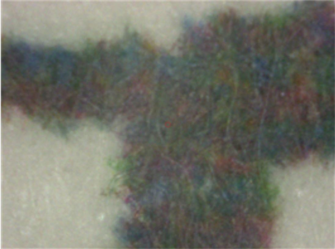
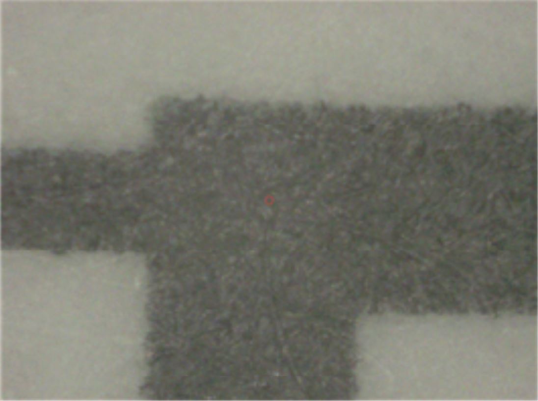
The quality difference between inkjet printing and offset printing ink was presented at a magnification of 100 times. The image produced by inkjet printing showed less defined lines due to ink spreading and bleeding (Fig. 2), while offset printing produced sharper and more defined lines and edges (Fig. 3). This suggested that offset printing provides better quality compared to inkjet printing, especially at a higher magnification.
Two popular printing techniques, i.e. inkjet and offset printing have their own strengths and weaknesses. Offset printing is well-known for producing sharp and crisp images due to its direct transfer method, which results in precise details and clean lines. On the other hand, inkjet printing sometimes struggles with ink diffusion and droplet size limitations, which may cause slightly less precise edges. Regarding color accuracy, offset printing is the winner, reproducing colors with great accuracy and precision consistently. However, inkjet printing has a wider color gamut, allowing it to produce more vibrant hues. Nevertheless, variations in ink and paper interactions may affect the color accuracy of inkjet printing.
Anionic inkjet inks and uncoated paper surfaces have the same charge characteristic, which makes it challenging to achieve optimal printing performance. This lack of electrostatic attraction can lead to problems like curling, wrinkling, and slow drying during the printing process. It is essential to rapidly immobilize the ink colorant on the paper surface while separating it from the carrier. However, uncontrolled ink absorption may cause excessive penetration into the paper, resulting in reduced optical density and color bleed. At the same time, insufficient absorption can lead to lateral spreading, edge raggedness, and line broadening. Striking a balance between rapid absorption and controlled ink migration is crucial.28)
3.4 Analyzing printing inks using XRF
Inkjet printing inks differ in inorganic components used depending on the printer company and type of ink, but in the case of the same ink, there are slightly different components depending on the manufacturing lot, but it is thought to be generally similar. Epson inkjet printer inks were separated and applied to the analysis paper and analyzed for the elemental composition of three colors. As shown in Table 2, the obvious difference in composition is the very high copper content of blue ink. The content of inorganic components in red or yellow ink is not high.
Blue ink used for inkjet printing contains higher copper content compared to red and yellow ink due to the use of copper-based pigments. Synthetic blue pigment, copper phthalocyanine (CuPC) blue, that replaced Prussian blue inks contains copper-based pigments.29) The result in Table 2 suggested that the higher copper content in blue ink used for inkjet printing was primarily due to the prevalent use of copper-based pigments.
Table 2.
Inkjet printing ink analysis by XRF
| Inorganic composition (%) | ||||||
| S | Ca | Mn | Fe | Cu | Zn | |
| Base Paper | n.d | 99.1 ± 0.01 | 0.15 ± 0.01 | 0.6 ± 0.01 | n.d | 0.01 |
| Red | 1.0 ± 1.5 | 94.7 ± 1.7 | 0.5 ± 0.2 | 1.6 ± 0.5 | 0.1 ± 0.1 | 0.7 ± 0.5 |
| Yellow | 0.1 ± 0.1 | 98.3 ± 0.5 | 0.2 ± 0.05 | 0.7 ± 0.1 | n.d | 0.7 ± 0.7 |
| Blue | 0.4 ± 0.2 | 51.2 ± 8.8 | 0.07 ± 0.001 | 0.25 ± 0.04 | 47.8 ± 8.6 | 0.2 ± 0.05 |
Table 3.
Black ink spot in paper analysis from different printing type inks by XRF
| Inorganic composition (%) | ||||||||
| Mg | Al | Si | S | Ca | Mn | Fe | Cu | |
| Inkjet printed *1 | 1.4 ± 0.1 | 0.15 ± 0.04 | 0.7 ± 0.01 | 0.7 ± 0.1 | 96.2 ± 0.1 | 0.2 ± 0.1 | 0.4 ± 0.1 | 0.2 ± 0.05 |
| offset printing *2 | 0.2 ± 0.1 | 1.4 ± 0.4 | 0.6 ± 0.1 | 1.0 ± 0.1 | 96.1 ± 0.3 | 0.2 ± 0.01 | 0.5 ± 0.1 | 0.1 ± 0.02 |
| Inkjet printed *2 | 37.6 ± 3.1 | 4.0 ± 1.2 | 19.4 ± 0.4 | 17.8 ± 2.5 | 4.2 ± 0.3 | 10.8 ± 1.1 | 6.2 ± 1.2 | |
| offset printing *2 | 5.2 ± 2.7 | 34.7 ± 6.9 | 14.7 ± 1.1 | 26.5 ± 5.9 | 5.0 ± 0.7 | 12.7 ± 3.4 | 1.2 ± 0.6 | |
Analysis of black ink spots from inkjet and offset printing showed some similarities and differences in their elemental compositions as shown in Table 3. Both inks mainly contained calcium, with inkjet printing containing 96.2% and offset printing containing 96.1%. This indicates that both printing methods use a common calcium-based pigment. The high calcium content in both inks suggests the use of calcium carbonate (CaCO3) as a common filler and extender in black inks. However, beyond the shared calcium dominance, the elemental composition of the ink spots diverged significantly. Inkjet printing resulted in a more diverse ink composition, incorporating magnesium (Mg), silicon (Si), sulfur (S), iron (Fe), and trace amounts of copper (Cu), manganese (Mn), and aluminum (Al). In contrast, offset printing yielded a simpler composition, primarily consisting of aluminum (Al), sulfur (S), silicon (Si), and iron (Fe), with magnesium, manganese, and copper present only as minor impurities.
The differences in elemental composition of ink on paper might be due to the ink formulations and printing processes used. Inkjet inks normally use water-based formulations with organic dyes or pigments, while offset printing uses oil-based inks with inorganic pigments. The choice of ink components and printing process parameters affect the final elemental composition of the ink on paper. Aqueous-based inks, which are the first generation inkjet inks, are still used today because they are environmentally friendly. They have minimal volatile organic compound (VOC) emissions and low toxicity. However, they have some drawbacks, such as relatively slow drying times on uncoated media and limited water resistance. Therefore, they are primarily used with coated media for indoor applications.30)
4. Conclusions
XRF analysis can distinguish between papers that use different fillers. Based on the content of magnesium in the filler, it was possible to determine whether it was ground calcium carbonate or precipitated calcium carbonate. Through micro-XRF analysis of the ink part of the printed paper, the inorganic components that make up the ink, and the printing style can be identified. Image analysis of the printed area by offset printing showed sharp images with precise details and clean lines. It also provides more color accuracy. On the other hand, inkjet print ink analysis and image analysis showed a wider color range with ink diffusion and droplet size limitations. Offset printing produced smoother results while inkjet printing might exhibit visible dot gain.
Additional research is necessary to investigate the impact of paper substrate on image quality. By integrating µ-XRF with other analytical techniques, it is possible to gain additional insights into the intricate interplay between paper, ink, and printing processes.


